501-918-3025
calsfoundation@cals.org
calsfoundation@cals.org
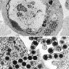
Top: Transmission electron micrograph (TEM) of a cell infected with ranaviruses, which gather in the cytoplasm and in the assembly bodies next to the contorted nucleus. Bottom: TEM of ranaviruses gathering close to the cell border where they can be seen leaving the cell via a process called budding.
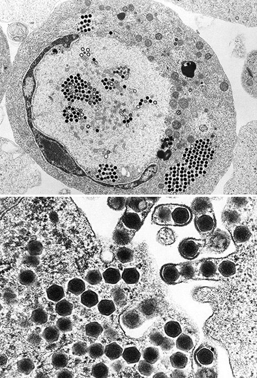
Courtesy of Electron Microscopy Unit, Australian Animal Health Laboratory


