calsfoundation@cals.org
Chytrid Fungus
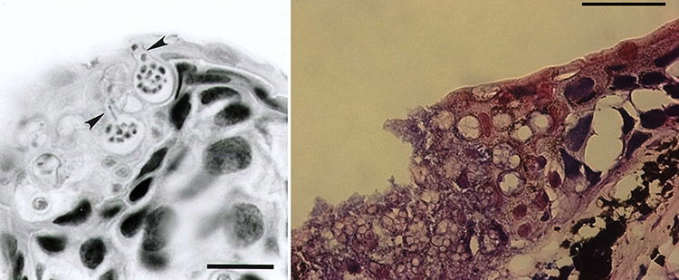
Photomicrographs of chytrid fungus in host skin biopsies. Left: View of Batrachochytrium dendrobatidis (Bd) sporangia in the skin of a Costa Rican variable harlequin toad (Atelopus varius); arrows indicate discharge tubes through which Bd zoospores exit the host cell; scale bar = 35 µm. Right: Bsal infection in the skin of a fire salamander (Salamandra salamandra), characterized by extensive epidermal necrosis, presence of high numbers of intra-epithelial colonial chytrid thalli, and loss of epithelial integrity; scale bar = 50 μm.


