501-918-3025
calsfoundation@cals.org
calsfoundation@cals.org
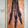
Snake fungal disease (ophidiomycosis) is an emerging infectious disease of numerous species of snakes caused by the fungus Ophidiomyces ophiodiicola within the family Onygenacea. Formerly, Ophidiomyces was classified as the Chrysoporium anamorph of Nannizziopsis vriesii species complex (CANV species complex), a group of fungi that are frequently associated with emerging infections in various groups of reptiles. However, recent phylogenetic analyses have demonstrated that CANV represents a species complex that also includes fungi of the genera Nannizziopsis and Paranannizziopsis and that Ophidiomyces is known to occur only on both colubrid and viperid snakes. It was first definitively identified in 2006 in a population of timber rattlesnakes (Crotalus horridus) in New Hampshire. However, although identification has not been conclusively proven because cultures or DNA analyses were not done, it is thought that the first documented case to represent ophidiomycosis was originally reported as “disseminated mycotic dermatitis” that actually occurred in a timber rattlesnake in October 1992 collected from near the Crater of Diamonds State Park in Pike County, Arkansas. The snake showed symptoms typical of ophidiomycosis, as it was noticeably emaciated and lethargic, and possessed numerous raised nodules. It was deposited as a voucher specimen in the Herpetological Collection at Arkansas State University in Jonesboro (Craighead County).
Ophidiomyces ophiodiicola is categorized as an environmental saprobe, that is, it normally feeds on decaying organic matter in the environment, and it is likely that the fungus resides in the soil. This is apparent due to its highly tolerant nature, as it can thrive in a wide range of pH from moderately acidic to highly basic (pH 5 to 11). It is also tolerant of drought and can utilize a number of complex compounds such as carbon, nitrogen, and sulfur. Considerable evidence that this fungus is a saprobe makes it likely that infection of snakes is opportunistic.
Early reports of snakes with skin infections of unknown origin have been irregular, but by 2020 O. ophiodiicola had been detected in snakes in at least thirty-eight U.S. states, one U.S. territory, and a single Canadian province. In addition to timber rattlesnakes, several other species of snakes are known to be afflicted with the disease, including black racers (Coluber constrictor), eastern indigos (Drymarchon couperi), milk snakes (Lampropeltis triangulum), western rat snakes (Pantherophis obsoletus), garter snakes (Thamnophis spp.), eastern massasauga (Sistrurus catenatus), and cottonmouths (Agkistrodon piscivorus). A new fungal clade, reported from European wild snakes, grows more slowly and is commonly associated with milder lesions. In captive snakes, the fungus has been reported globally. However, researchers suspect that ophidiomycosis might be more widely distributed than these documented cases seem to suggest, because there are limited efforts to monitor the health of many snake populations. Therefore, this disease may also be underreported in populations where it affects snakes infrequently or in species that develop less severe pathology.
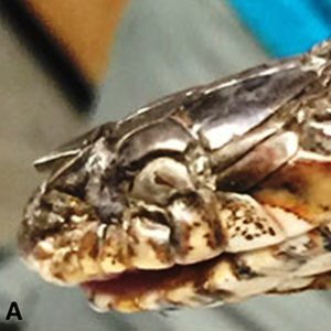
Signs of ophidiomycosis include localized thickening or crusting of the skin, ulcerated skin, abnormal bumps or nodules under the skin, abnormal molting with white opaque cloudiness of the eyes, and facial disfiguration that can be quite severe, potentially leading to emaciation and, ultimately, death. Many snake populations are already in decline, and the recent emergence of this fungus may accelerate this decline, causing certain populations to vanish entirely from some locations.
In 2016, several scientists published a study demonstrating that ophidiomycosis is widespread in eastern North America, has a broad host range among snakes, and is the predominant cause of skin infections in wild snakes. The study also showed that O. ophiodiicola frequently causes non-lethal infections in snakes and that environmental changes are likely causing the recent emergence of severe and fatal infections in some snake populations. Indeed, several studies have indicated that temperature is a significant factor affecting the growth of O. ophiodiicola. Data also suggests that with increasing global temperatures due to climate change, snake populations will be more vulnerable to O. ophiodiicola. This portends that snake populations hibernating or in winter dormancy in the lower thermal range of 0 to 10 degrees C (32 to 50 degrees F) should have a reduced infection during the spring and summer compared to snakes that hibernate in the upper thermal range > 10 C (> 42 F).
One of the characteristic clinical signs of the disease is facial swelling. It can progress internally from the nasal cavity via the eyes, throat, and lungs, potentially causing eye infections and pneumonia. Externally, the fungus additionally spreads along the neck, body, and tail, forming scattered nodules (lumps and bumps) or ulcerations. In experimental infections, the incubation period averages between 30 to 37 days with some showing clinical signs as early as day 12 of inoculation. In rare cases in which there are injuries secondary to the infection, the fungus is capable of penetrating the body, causing a systemic fungal infection resulting in nodules on the coelomic cavity fat pad, kidneys, liver, and alveoli of lungs. Experimental data has shown that infected snakes surviving an average of 90 days have a 40 percent mortality rate.
Interestingly, O. ophiodiicola can be shed into the environment by infected snakes and spread from the environment to other snakes, particularly to those that share dens during winter dormancy. However, there is no conclusive evidence of snake-to-snake transmission. When humans track contaminated soil imbedded in clothing or shoes, they may spread the fungus to new locations. Not all infected snakes succumb to the disease, as two infected C. horridus (Viperidae) had improved health over ten weeks in captivity without treatment to the point of one being asymptomatic. Treatment with antifungal medications has not been successful in colubrid snakes.
Ophidiomycosis is diagnosed by identification of the typical macroscopic skin lesions as well as laboratory identification of the fungus via culture, histopathological examination of suspected tissues (skin biopsies), and real-time or quantitative polymerase chain reaction (rtPCR and qPCR).
For additional information:
Allender, Matthew C., M. Dreslik, S. Wylie, C. Phillips, D. Wylie, C. Maddox, M. A. Delaney, and M. J. Kinsel. “An Unusual Mortality Event Associated with Chrysosporium in Eastern Massasauga Rattlesnakes (Sistrurus catenatus catenatus).” Emerging Infectious Diseases 17 (2011): 2383–2384.
Allender, Matthew C., D. B. Raudabaugh, F. H. Gleason, and A. N. Miller. “The Natural History, Ecology, and Epidemiology of Ophidiomyces ophiodiicola and its Potential Impact on Free-Ranging Snakes Populations.” Fungal Ecology 17 (2015): 187–196.
Allender, Matthew C., D. Bunick, E. Dzhaman, L. Burrus, and C. Maddox. “Development and Use of Real-Time Polymerase Chain Reaction Assay for Detection of Ophidiomyces ophiodiicola in Snakes.” Journal of Veterinary Diagnostics and Investigation 27 (2015): 217–220.
Allender, Matthew C., S. Baker, D. Wylie, D. Loper, M. J. Dreslik, C. A. Phillips, C. Maddox, and E. A. Driskell. “Development of Snake Fungal Disease after Experimental Challenge with Ophidiomyces ophiodiicola in Cottonmouths (Agkistrodon piscivorous).” PLoS One 10 (2015): e0140193.
Baker, Sarah, Ellen Haynes, Kristin Stanford, Michelle Christman, Kenneth Conley, Salvatore Frasca Jr., Robert J. Ossiboff, Denae Lobato, and Matthew C. Allender. “Case Definition and Diagnostic Testing for Snake Fungal Disease.” Herpetological Review 50 (2018): 279‒285.
Barber, Diane M., Vicky A. Poole, Carlos R. Sanchez, Patrick Roady, and Matthew C. Allender. “Snake Fungal Infection Associated with Fusarium Found in Nerodia erythrogaster transversa (Blotched Water Snake) in Texas, USA.” Herpetological Review 47 (2015): 39‒42.
Cheatwood, J. L., Eliott R. Jacobson, P. G. May, T. M. Farrell, B. L. Homer, D. A. Samuelson, and J. W. Kimbrough. “An Outbreak of Fungal Dermatitis and Stomatitis in a Free-Ranging Population of Pigmy Rattlesnakes (Sistrurus miliarius barbouri) in Florida.” Journal of Wildlife Diseases 39 (2003): 329–337.
Dolinski, A. C., Matthew C. Allender, V. Hsiao, and C. W. Maddox. “Systemic Ophidiomyces ophiodiicola Infection in a Free-Ranging Plains Garter Snake (Thamnophis radix).” Journal of Herpetological Medicine and Surgery 24 (2014): 7–10.
Franklinos, L. H., J. M. Lorch, E. Bohuski, J. R. Fernandez, O. N. Wright, L. Fitzpatrick, S. Petrovan, C. Durrant, C. Linton, V. Baláž, and A. A. Cunningham AA. “Emerging Fungal Pathogen Ophidiomyces ophiodiicola in Wild European Snakes.” Scientific Reports 7 (2017): 3844.
Glorioso, Brad M., James H. Waddle, E. Earl Green, and Jeffery M. Lorch. “First Documented Case of Snake Fungal Disease in a Free-Ranging Wild Snake in Louisiana.” Southeastern Naturalist 15: N4‒N6.
Jacobson, Elliott R. Infectious Diseases and Pathology of Reptiles: Color Atlas and Text. Boca Raton, FL: CRC Press, 2007.
Januszkiewicz, Eric, Nicole Chinnici, and Thomas C. LaDuke. “Detection of Snake Fungal Disease Caused by Ophidiomyces ophiodiicola Among Timber Rattlesnake (Crotalus horridus) in Pennsylvania, USA.” Herpetological Review 50: 286‒288.
Lorch, Jeffrey M., Julia Lankton, Katrien Werner, Elizabeth A. Falendysz, Kevin McCurley, and David S. Blehert. “Experimental Infection of Snakes with Ophidiomyces ophiodiicola Causes Pathological Changes That Typify Snake Fungal Disease.” MBio 6 (2015): e01534-15.
McAllister, Chris T., Stephen R. Goldberg, H. J. Holshuh, and Stanley E. Trauth. “Disseminated Mycotic Dermatitis in a Wild-Caught Timber Rattlesnake, Crotalus horridus (Serpentes: Viperidae), from Arkansas.” Texas Journal of Science 45 (1993): 279‒281.
McBride, M. P., K. B. Wojick, T. A. Georoff, J. Kimbro, M. M. Garner, X. Wang, A. L. Childress, and J. F. X. Wellehan. “Ophidiomyces ophiodiicola Dermatitis in Eight Free-Ranging Timber Rattlesnakes (Crotalus horridus) from Massachusetts.” Journal of Zoo and Wildlife Medicine 46 (2015): 86–94.
Rajeev, S., D. A. Sutton, B. L. Wickes, D. L. Miller, D. Giri, M. Van Meter, E. H. Thompson, M. G. Rinaldi, A. M. Romanelli, J. F. Cano, and J. Guarro. “Isolation and Characterization of a New Fungal Species, Chrysosporium ophiodiicola, from a Mycotic Granuloma of a Black Rat Snake (Elaphe obsoleta obsoleta).” Journal of Clinical Microbiology 47 (2009): 1264–1268.
Tetzlaff, S., Matthew Allender, M. Ravesi, J. Smith, and B. Kingsbury. “First Report of Snake Fungal Disease from Michigan, USA Involving Massasaugas, Sistrurus catenatus (Rafinesque 1818).” Herpetology Notes 8 (2015): 31–33.
Chris T. McAllister
Eastern Oklahoma State College


Comments
No comments on this entry yet.