501-918-3025
calsfoundation@cals.org
calsfoundation@cals.org
A lichen is a composite organism that is the result of a simple photosynthesizing organism (green algae or cyanobacteria/blue-green algae) living among filaments (hyphae) of multiple fungi species in a mutually beneficial symbiotic relationship. Although lichens had been recognized as organisms for many years, it was not until 1867, when Swiss botanist Simon Schwendener (1829‒1919) proposed his dual theory of lichens, that the true nature of the lichen association began to emerge. The green algae or cyanobacteria benefit by being protected from the environment by the filaments of the fungi, which also gather moisture and nutrients from the environment, and (usually) provide an anchor to it, whereas the fungi benefit from the carbohydrates produced by the green algae or cyanobacteria via photosynthesis. About 250 species of lichens can be found in Arkansas.
Lichens independently emerged from fungi associating with algae and cyanobacteria multiple times throughout history. In the past, some lichen specialists placed them in their own division (Mycophycophyta); however, this taxonomic placing is no longer accepted because the components belong to separate lineages. Indeed, neither the ascolichens nor the basidiolichens form monophyletic lineages in their respective fungal phyla, but they do form several major solely or primarily lichen-forming groups within each phylum.
The fossil record of lichens is very poor. This is because lichens do not ordinarily produce fossils, as the extreme habitats in which they dominate do not produce useful specimens. However, there are fossilized lichens embedded in amber. The fossilized Anzia from northern Europe found in pieces of amber dates back to the early Tertiary, approximately 40 million years ago. Dating to the early to middle Miocene, lichen fragments are known from fossil leaf beds, such as lung wort, Lobaria from northern California. Dating from the early Devonian, about 400 million years ago, is the oldest fossil lichen in which both symbiotic partners have been recovered.
There are about 13,500 to 17,000 known species of lichens, possessing many colors, sizes, and forms. It is estimated that about six percent of Earth’s terrestrial surface is covered by lichens, and nearly twenty percent of known fungal species are associated with them. Within the division Ascomycota is the largest number of lichenized fungal species, with about forty percent of species forming such an association. Some of these lichenized fungi occur in orders with non-lichenized fungi that live as saprotrophs (organisms that feed on detritus) or as plant parasites. Other lichen fungi occur in only five orders (Graphidales, Gyalectales, Peltigerales, Pertusariales, and Teloschistales) in which all members utilize these habits. Overall, about ninety-eight percent of lichens have an ascomycetous mycobiont. Next to the Ascomycota, the greatest number of lichenized fungi belong in the unassigned fungi imperfecti, a catch-all taxonomic category for fungi whose sexual form of reproduction has never been observed. Comparatively few occur in the division Basidiomycetes (mostly agarics) that are lichenized.
Depending on the species, lichen associations may be examples of mutualism, commensalism, or even parasitism. In fact, there is sufficient support to suggest that the lichen symbiosis is parasitic or commensalistic, rather than mutualistic. The photosynthetic partner can exist in nature independently of the fungal partner, but not vice versa. However, lichens are excellent examples of successful symbiosis as evidenced by the fact that lichens can be found in almost every habitat and geographic region on the planet. For example, two species within two genera of green algae are found in over thirty-five percent of all lichens but can only rarely be found living on their own outside of one. In addition, the fungus Geosiphon pyriforme (Glomeromycota) is unique in that it encloses a cyanobacterial symbiont (Nostoc) inside its cells. Geosiphon is not usually considered a true lichen, and its unique symbiosis was not recognized for many years. Fungi from the order Verrucariales also form marine lichens with the unusual photobiont brown algae Petroderma maculiforme and have a symbiotic relationship with seaweed (rockweed) and Blidingia minima, where the algae are the dominant components. When exposed to air, the fungus is thought to help the rockweeds resist desiccation. In addition, lichens can also use psychrophilic yellow-green algae (Heterococcus) as their symbiotic partner.
Although some photosynthetic associates in lichens may survive outside the lichen, the lichen symbiotic association extends the ecological range of both partners, whereby most descriptions of lichen associations describe them as symbiotic. While symbiotic, however, the relationship is probably not mutualistic, given that the algae give up a disproportionate amount of their carbohydrates. In fact, the algae may lose up to eight percent of their sugar production to the fungus. However, both associates gain water and mineral nutrients mainly from the atmosphere, through rain and dust particles. By retaining water, the fungal partner protects the alga by serving as a larger capture area for mineral nutrients and, in some cases, provides minerals obtained from the substrate. If a cyanobacterium is present, as a primary partner or another symbiont in addition to a green alga as in certain tripartite lichens, they can fix atmospheric nitrogen, complementing the physiologies of the green alga.
Unlike plants, lichens do not possess roots that absorb nutrients and water, but similarly via photosynthesis, they produce their own nutrition. When they grow on plants, they use the plants as a substrate and not as parasites. The algal or cyanobacterial cells are photosynthetic and, as in plants, they reduce atmospheric CO2 into organic carbon sugars to feed both symbionts.
Lichens are distributed across the biosphere from sea level to high alpine elevations, in many environmental conditions, and they can grow on almost any surface. Lichens are considered to be among the oldest living organisms. After a catastrophic event such as a landslide, they are among the first biota to grow on exposed fresh rock. The long lifespan and slow and regular growth rate of some lichens on exposed rock surfaces can even be used to date events (termed lichenometry). As lichens grow at a near constant rate, they are useful in dating objects that are relatively undisturbed. This was a technique that has found many applications and was introduced in the 1950s by Austrian botanist Roland Beschel (1928‒1971). It determines the age of exposed rock by using the presumed regular but slow rate of lichen growth. As radiocarbon dating techniques are less accurate over this period, the technique is especially useful for dating surfaces less than 500 years old. For example, it has been used to date gravestones in Arkansas at the Cedar Grove Cemetery near Clarksville (Johnson County).
Lichens are capable of growing on and in a wide range of substrates and habitats, including some of the most extreme conditions on Earth, including arctic tundra, hot xeric deserts, rocky coasts, and even on toxic slag heaps from mining operations. They can live inside solid rock, growing between the grains, and in the soil as part of a biological soil crust in arid habitats such as deserts. Some lichens do not grow on anything at all, however, living out their lifetimes blowing throughout the environment. Lichens are especially common as composites growing on bark, leaves, mosses, and even on other lichens and hanging from branches, such as epiphytes in tropical rain forests and in temperate woodland. Lichens also grow on rocks, walls, gravestones, roofs, and exposed soil surfaces, as well as in the soil as part of a biological soil crust.
Lichens grow in a wide range of morphologies. The shape is usually determined by the fungal filament organization. The vegetative body parts (non-reproductive tissues) are called the thallus. Since the thallus is usually the most visually prominent part of the specimen, they are typically grouped by thallus type. Thallus growth forms typically correspond to a few basic internal structure types. The filaments (hyphae) grow by branching then rejoining to create a dense or loose anastomose mesh. Generally, the fungal mesh surrounds the algal or cyanobacterial cells, often enclosing them within complex fungal tissues that are unique to lichen associations. The thallus may or may not have a protective top layer (skin) of densely packed fungal filaments, often containing a second fungal species, which is called a cortex. The cortex is made of tightly woven, densely packed, and agglutinated (glued together) fungal filaments. The dense packing makes the cortex act like a protective layer, keeping other organisms out and reducing the intensity of sunlight on the layers below.
Immediately beneath the cortical layer is the photobiontic layer (or symbiont) layer. This layer has less densely packed fungal filaments, with the photosynthetic associate embedded in them. Each cell or group of cells of the photobiont is usually individually wrapped by hyphae, and in some cases penetrated by a highly modified rootlike structure called a haustorium.
The medulla is the lowest layer just beneath the symbiont layer. It is less densely packed with fungal filaments than the layers above. Root-like fungal multicellular structures called rhizines usually grow from the lower cortex to attach or anchor the lichen to the substrate. The medulla may form a cottony white inner core for the branchlike thallus, or it may be hollow.
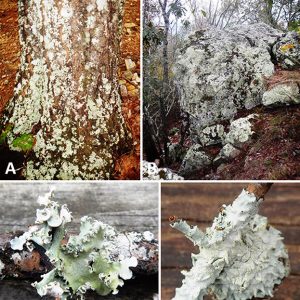
Lichens come in several lichen thallus growth forms as follows: (1) those growing like a tuft or multiple-branched leafless mini-shrub, upright or hanging down, three-dimensional branches with nearly round cross section (terete) or flattened are termed fruticose; (2) those growing in two-dimensional, flat, leaf-like lobes are called foliose; (3) those with crust-like flakes that lie on the surface are crustose; (4) those with a powdery-like appearance are leprose; (5) those having jelly-like forms or when dry and leafy are gelatinous; (6) those formed of small leaf-like scales, crustose below, but free at the tips or where the edges lift up are squamulose; (7) those that are stringy, similar to matted hair, are filamentous; (8) those that are wispy, similar to teased wool, are byssoid; and (9) those having none of the above forms are called structureless. Lichens that are either bush-like or leafy are macrolichens and have more than one species of fungus in their tissues, and all other lichens are termed microlichens.
Morphological structures involved in reproduction often appear as bumps, discs, or squiggly lines on the surface of the thallus. The most noticeable reproductive parts are often circular, raised, plate-like or disc-like outgrowths, with furrowed edges. Some lichens that are dramatic in color or appearance can grow inside solid rock between the grains (endolithic lichens), with only the sexual fruiting part visible growing outside the rock. Forms of these sexual parts are not in the above growth form categories.
Lichens come in many colors, usually determined by the photosynthetic component. Yellow usnic acid (a special pigment and antimicrobial compound found almost exclusively in lichens) gives lichens a variety of colors, including yellow, brown, red, and orange, especially in exposed, xeric habitats. When special pigments are absent, lichens are usually bright green to olive gray when wet, whereas when dry, they are gray or grayish-green to brown. Moisture causes the cortex (surface skin) to become more transparent, exposing the green photobiont layer. Interestingly, a spectacular color display can occur when different-colored lichens cover large areas of exposed rock surfaces, or when lichens cover or hang from bark following rain, when the patches of diverse colors appear to come to life or glow in brilliant displays. A good example is found in Yosemite National Park, where colonies of lichens cover the vast rock faces of the park. However, lichens can sometimes be a nuisance, such as an ongoing lichen overgrowth problem on Mount Rushmore National Memorial in South Dakota that requires mountain-climbing conservators to periodically clean the monument.
Lichens are pioneer species and among the first living organisms to grow on bare rock or areas denuded of life after a catastrophe. Lichens are also particularly important to conditions in wet montane temperate forests, where they contribute significantly to epiphytic biomass, which, before the 1980 stratovolcanic eruption, was widespread at Mount St. Helens in Washington State. They have also been widely used as environmental indicators or bio-indicators. In terms of perturbations, several lichens are very sensitive to environmental disturbances and have been used to assess air pollution, ozone depletion, and heavy metal contamination. This interaction between lichens and air pollution has been used as a means of monitoring air quality since 1859, with more systematic methods developed in 1866 by Finnish naturalist William Nylander (1822‒1899). He observed that some lichen species present within Luxembourg Gardens, in Paris, France, were absent in other parts of the city. He attributed these differences to air quality. Over the next thirty years, fumes from coal-burning industrial furnaces gradually led to the eradication of the lichen population within the park. For example, if the atmosphere is very badly polluted with sulphur dioxide (SO2), none may be present, and only green algae may be found. On the other hand, shrubby, hairy, and leafy lichens become abundant in clean air. In urban areas, a few lichen species can tolerate quite high levels of pollution and are commonly found on pavement, walls, and tree bark. Since industrialization, many of the shrubby and leafy lichens such as Lobaria, Ramalina, and Usnea species have very limited ranges, often being confined to the parts with the purest air.
Lichens have been used in making dyes, perfumes, and traditional medicines. Many lichens also produce secondary compounds, including pigments that reduce harmful amounts of sunlight and powerful toxins that reduce herbivory or kill bacteria. Lichens also have potential diagnostic or therapeutic value as broad-spectrum antibiotics, while a few are associated respectively to antiseptic similarities.
A few species of lichens are eaten by arthropods or grazing mammals, such as reindeer living in Arctic regions. Also, the larvae of a number of lepidopteran (certain arctiid moths) species feed exclusively on lichens. However, lichens are very low in protein but high in carbohydrates, making them unsuitable as food for some animals.
For humans, lichens are eaten by many different cultures across the globe. Although some lichens are only eaten in times of starvation, others are a staple food or even a delicacy. Iceland moss (Cetraria islandica) was an important food source for the peoples of northern Europe and was eaten as a bread, porridge, pudding, salad, or soup. Another type, wila or Fremont’s horsehair lichen (Bryoria fremontii), was an important food in parts of North America for many Native Americans, where it was usually pit-cooked. The northern peoples of North America and Siberia traditionally eat the partially digested reindeer lichen (Cladina spp.) after they remove it from the rumen of caribou (Rangifer tarandus) that have been killed. Rock tripe (Umbilicaria spp. and Lasalia spp.) are lichens that have frequently been used as an emergency food in North America, and one species, Umbilicaria esculenta, generally found at high altitudes on mountain rocks in East Asia, is used in a variety of traditional Asian (Japanese and Korean) cuisine and a restorative medicine in traditional Chinese medicine. It has been shown to inhibit cholesterol synthesis, work as an anti-inflammatory, and also have anti-tumor properties. However, two impediments are often encountered when humans eat lichens: (1) polysaccharides in lichens are generally indigestible, and (2) lichens usually contain mildly toxic secondary compounds that should be removed before eating. Although very few lichens (mostly yellow in color) are poisonous, those high in vulpinic acid (C19H14O5) or usnic acid (C18H16O7) are toxic.
Lichens often have a regular but very slow growth rate of less than one mm (0.04 in.) per year. For example, most crustose lichens grow only 1 to 2 mm (0.04 to 0.08 in.) in diameter per year. Lichens may have long life spans, with some considered to be among the oldest living organisms. Indeed, an Arctic species commonly called the map lichen (Rhizocarpon geographicum) has been dated at 8,600 years, apparently the world’s oldest living organism. It is also broadly distributed over the world and may be found in most cold areas with exposed rock surfaces.
Interestingly, the European Space Agency (an intergovernmental organization dedicated to the exploration of space) has reported that lichens can survive unprotected in outer space. In one experiment, two species (Rhizocarpon geographicum and Xanthoria elegans) were sealed in a capsule and launched in 2005 on a Russian Soyuz rocket. Once in orbit, their capsules were opened and the lichens were exposed directly to the vacuum of outer space with its unstable temperatures and cosmic radiation. Sixteen days later, the lichens were returned to Earth and were found to be unchanged in their physiology, particularly in their ability to photosynthesize.
Many lichens reproduce asexually, either by a piece breaking off and growing on its own (vegetative reproduction) or through the dispersal of lichen propagules (diaspores) containing a few algal cells surrounded by fungal cells. On the other hand, structures involved in sexual reproduction often appear as discs, bumps, or squiggly lines on the surface of the thallus. Only the fungal partner within the lichen reproduces sexually. Like other fungi, many lichen fungi reproduce sexually, producing spores formed by meiosis and fusion of gametes. Following dispersal, such fungal spores must meet with a compatible algal partner before any functional composite lichen can form.
The initial publications on lichens in North America included only a few references to these organisms from Arkansas. The earliest of these was published in 1882 and listed three species from the state. Several decades later, a publication from 1935 listed a total of five species from Arkansas (three of the same from 1882), and in 1941 another eighteen specimens including the largest genus of foliose lichens (Parmelia) collected in Newton County. There were also collections of cup lichens (Cladogonia) from Arkansas in the 1940s. A checklist of species in 1945 included fifty-four species mostly collected from Conway, Drew, and Pulaski counties. Various publications between 1955 and 1967 listed a total of sixty-two species. This included the type specimen of Parmelia (=Xanthoparmelia) hypomelaena from near Malvern (Hot Spring County). Various monographs on lichen flora published between 1963 and 1973 have included Arkansas specimens of Leptogium, Loharia, Ochrolechia, and Physcia. In 1979, a key to the foliose and fruticose lichens of the United States added about 100 species to the state checklist. By 2021, about 250 species of lichens were thought to occur in the state. However, additional research is sorely needed to continue looking for and describing new lichens in Arkansas.
For additional information:
Bustinza, F. “Antibacterial Substances from Lichens.” Economic Botany 6 (1952):402–406.
Fink, Bruce. “Lichen Flora of United States.” Ann Arbor: University of Michigan Press, 1935.
Gargas, A., P. T. Depriest, M. Grube, and A. Tehler. “Multiple Origins of Lichen Symbioses in Fungi Suggested by SSU rDNA Phylogeny.” Science 268 (1995): 1492–1495.
Grube, M., M. Cardinale, J. V. De Castro Jr., H. Müller, and G. Berg. “Species-Specific Structural and Functional Diversity of Bacterial Communities in Lichen Symbioses.” The ISME Journal 3 (2009): 1105–1115.
Hagiwara, K., and P. R. Wright, et al. “Comparative Analysis of the Antioxidant Properties of Icelandic and Hawaiian Lichens.” Environmental Microbiology 18 (2015): 2319–2325.
Hale, Mason E., Jr. How to Know the Lichens, 2nd ed. Dubuque, IA: William C. Brown Co., 1979.
Hawksworth, D. L. “The Variety of Fungal-Algal Symbioses, their Evolutionary Significance, and the Nature of Lichens.” Botanical Journal of the Linnean Society 96 (1988): 3–20.
Herre, Albert, W. C. T. “Some Arkansas Lichens.” The Bryologist 48 (1945):85‒90.
Johnson, George T. “Three Species of Trypethelium New to Arkansas.” Proceedings of the Arkansas Academy of Science 33 (1979): 82‒83. https://scholarworks.uark.edu/cgi/viewcontent.cgi?article=2718&context=jaas (accessed February 24, 2021).
Kluthe, Brandy Garrett, Margaret Guiccioni, and Steven L. Stephenson. “Using Lichenometry, Dendrochronology, and Historical Data to Establish the Relative Age of an Abandoned Cemetery in Northern Arkansas.” Ethnobiology Letters 9 (2018): 253–262.
Lendemer, J. C. “A Taxonomic Revision of the North American Species of Lepraria s. l. that Produce Divaricatic Acid, with Notes on the Type Species of the Genus L. incana.” Mycologia 103 (2011): 1216–1229.
Lutzoni, Francois, F. Kauff, C. J. Cox, D. McLaughlin, G. Celio, B. Dentinger, M. Padamsee, and D. Hibbett, et al. “Assembling the Fungal Tree of Life: Progress, Classification, and Evolution of Subcellular Traits.” American Journal of Botany 91 (2004): 1446–1480.
Lutzoni, Francois, Mark Pagel, and Valerie Reeb. “Major Fungal Lineages are Derived from Lichen Symbiotic Ancestors.” Nature 411 (2001): 937–940.
Margulis, Lynn, and Eva Barreno. “Looking at Lichens.” BioScience 53 (2003): 776.
McCarroll, D. “Lichens: Lichenometric Dating of Diachronous Surfaces.” Earth Surface Processes and Landforms 20 (1995): 829–831.
Moore, Jewel E. Checklist of the Lichens of Arkansas. Arkansas Academy of Science, Arkansas Biota Survey Checklist No. 30, 1981.
———. “Lichens of Arkansas I: A Summary of Current Information.” Proceedings of the Arkansas Academy of Science 33 (1979): 85–87. Online at https://scholarworks.uark.edu/cgi/viewcontent.cgi?article=2721&context=jaas (accessed February 24, 2021).
———. “Lichens of Arkansas II: Additional State Records through Computer Search.” Journal of the Arkansas Academy of Science 35 (1981): 88. Online at https://scholarworks.uark.edu/cgi/viewcontent.cgi?article=2627&context=jaas (accessed February 24, 2021).
———. “A Study of the Vegetation of Petit Jean Mountain in Central Arkansas.” Castanea 30 (1965): 1‒37.
Moore, Jewel E., and Steve L. Timme. “Bryophyte-lichen Communities Within Hot Springs National Park, Arkansas I.” Proceedings of the Arkansas Academy of Science 37 (1983): 96. Online at https://scholarworks.uark.edu/cgi/viewcontent.cgi?article=2547&context=jaas (accessed February 24, 2021).
Muller, G. “Lichenometry and Environmental History.” Environmental History 11 (2006): 604–609.
Oksanen, I. “Ecological and Biotechnological Aspects of Lichens.” Applied Microbiology and Biotechnology 73 (2006): 723–734.
Taylor, T. N., H. Hass, W. Remy, and H. Kerp. “The Oldest Fossil Lichen.” Nature 378 (1995): 244.
Tuckerman, Edward. Synopsis of North American Lichens. Part I. Collected Lichenological Papers in University of Central Arkansas Library, Conway, Arkansas, 1882.
Chris T. McAllister
Eastern Oklahoma State College


Comments
No comments on this entry yet.