501-918-3025
calsfoundation@cals.org
calsfoundation@cals.org
The phylum Porifera, which contains the sponges, is a highly successful group of metazoan animals that includes about 8,600 living species of marine and freshwater forms as well as some that inhabit brackish waters. The majority are marine, but there are about 150 species of freshwater sponges, including twenty-seven to thirty species found in North America north of Mexico. The family Spongillidae is the most speciose and widespread group of freshwater sponges and includes twenty-two genera and more than 130 species from a wide variety of habitats. Seven species of freshwater sponges have been documented in Arkansas.
Sponges are an ancient group of asymmetrical invertebrates with a fossil record preceding the early Cambrian period (541 million years ago), and even today they are the simplest multicellular animals. Generally, the body of a sponge is an assemblage of cells embedded in an extracellular matrix and supported by a skeleton of minute needle-like spicules and proteins. They have a unique water-current system with various degrees of complexity. Because of their simple body plan, zoologists did not completely recognize sponges as animals until well into the nineteenth century, as they were previously thought to be plants. Molecular analysis clearly showed that sponges are phylogenetically grouped with animals. They are paraphyletic because the calcareous “sponges” are more closely related to other animal taxa than they are to siliceous sponges.
Sponges vary from a few millimeters to over two meters (6.6 ft.) in diameter. Some sponges may live to be 2,300 years old based on recent studies of size and growth in the Caribbean reef sponge, Xestospongia muta. Most marine sponges are brightly colored due to pigments in their dermal cells. Common coloration of sponges are those of the rainbow such as yellow, orange, green, red, and purple. Freshwater sponges tend to be more sedate and not especially colorful.
The classification of sponges is based on spicule form and chemical composition. Living sponges have traditionally been divided into three or perhaps four classes: Homoscleromorpha, Calcispongiae, Hexactinellida, and Demospongiae. The former is considered a clade with an absent skeleton or a skeleton of siliceous spicules without an axial filament. Species of the Calcispongiae (Calcarea) typically have spicules of calcium carbonate with one, three, or four rays. Glass sponges make up the Hexactinellida (Hyalospongiae) and have six-rayed siliceous spicules and a syncytial trabecular reticulum, and members of Demospongiae (Demospongia) have spicules not in six rays and a skeleton of siliceous spicules that develop around an axial filament, or spongin fibers, or both.
Homoscleromorpha are considered by some to be a subclass rather than class of sponges. Formerly, these sponges were placed in Demospongiae. They are marine sponges that live in cryptic habitats that possess unique features such as a pinacoderm layer and a true basement membrane. They include the genera Corticium, Oscarella, and Plakina.
Calcispongiae are calcareous sponges because their spicules are composed of calcium carbonate. Spicules are straight or have three or four rays. They tend to be small, 10 cm (3.9 in.) or less in height, and tubular or vase-shaped. They may be asconoid, syconoid, or leuconoid in body plan. In zoology labs, students usually study Clathrina, Leucosolenia and Sycon (often called Scypha or Grantia by biological supply companies).
Hexactinellida are the glass sponges. Basically, all glass sponges are marine, deep-water forms that are rarely seen. Most are superficially radially symmetrical, with vase- or funnel-shaped bodies attached to the substrate by stalks of root spicules. Size ranges from 7.5 cm (3.0 in.) to more than 1.3 m (4.3 ft.) in length. They possess six-rayed (triaxon) siliceous spicules that are commonly formed together into a network creating a glass-like or skeletal structure. A classic and most famous example of a glass sponge is Euplectella or Venus’s flower basket.
Demospongiae contains perhaps 90 percent of all living sponges, including all the large sponges and those used in industry as well as in the home. Spicules in the group are siliceous, but not six-rayed, and are grouped as either microscleres or megascleres. Spicules may be bound together by spongin (a form of collagen) or are absent. Bath sponges (Spongia and Hippospongia) are members of this group. Bath sponges have spongin skeletons and lack siliceous spicules altogether. All members of this class have a leuconoid body plan and most are marine, except the members of the freshwater family Spongillidae. Examples include the genera Hippospongia, Meyenia, Spongia, and Spongilla.
There are four main types of cells in sponges: pinacocytes, porocytes, choanocytes, and archeocytes (amoebocytes or mesenchyme cells). The pinacocytes are the cells of the external epithelium that function in contraction and structure. The porocytes are the cells that form pores that function to allow flow of water. The choanocytes line the flagellated canals and chambers and mainly function to create water flow. The archaeocytes are amoeboid cells that can be non-sessile and found in cellular matrix. Their main functions are digestion and to secrete structural components, including spicules and spongin.
Most animals generally are mobile; however, adult sponges (larvae are motile) are sessile (i.e., they are attached to a substrate and do not physically move around). Instead, they filter water that is drawn into a central cavity in the body. The ostia (tiny dermal pores) through which the water enters give rise to their name, Porifera (“pore bearing”). Unique specialized cells known as choanocytes move water into the sponge in an aquiferous system. Each choanocyte (in contact with an amoebocyte) has a tiny flagellum that draws water, bringing food and oxygen past each cell, as well as carrying away waste products through oscula (osculum). Thus, the sponge body is an efficient aquatic filter for removing suspended particles from the water.
Sponges are always attached to some type of substrate such as a rock, shell, coral, stick, or other submerged object. Some growth forms of sponges vary from erect, branched, or lobed, while others are low encrusting forms. Large sponges tend to harbor a great variety of invertebrate commensal organisms. Many sponges grow on other living animals such as mollusks, barnacles, brachiopods, corals, or hydroids.
Interestingly, the skeletal framework of a sponge can be fibrous and/or rigid. A rigid skeleton consists of calcareous or siliceous support structures known as spicules. Alternatively, the fibrous portion of the skeleton comes from collagen protein fibrils contained in the intercellular matrix of all sponges. Collagen comes in a number of types that differ in chemical composition and form (e.g., fibers, filaments, or masses surrounding spicules). Spongin is a well-known form. Sponges often contain microalgae and cyanobacteria (blue-green algae) on the body surface and inside the body itself.
As stated previously, sponges feed by collecting suspended particles from water drawn into the sponge through internal canal systems. Water enters the sponge through small incurrent pores, called dermal ostia, which have an average diameter of 50 micrometers (µm). Once inside the body, water is directed past the choanocytes where food particles (detritus, planktonic organisms) are collected on the choanocyte collar. Some large sponges can filter up to 1,500 liters (396 gallons) of water per day. Sponges feed by phagocytosis, whereby the food is taken into the choanocytes. There are three main designs for a sponge body regarding how water flows in the animal.
Most sponges can be separated based on their type of canal system. The more complex a sponge condition, the more particles it can filter from the water column. The simplest body plan is called the asconoid system wherein the choanocytes lie in a large chamber called the spongocoel (atrium) where water has moved there through the ostia. Asconoid sponges like Leucosolenia are tube shaped and rarely exceed 10 cm (3.9 in.) in length, occurring only in the class Calcispongiae. In the second system, the syconoid, the choanocytes lie in canals. Syconoid sponges like Sycon are larger and sometimes look like large asconoids in that they have a tubular body and single osculum, the exit opening from the spongocoel. In the syconoid system, water enters the body through dermal ostia that lead to incurrent canals. The water then passes through tiny openings called prosopyles, into the radial canals where food is ingested by the lined choanocytes. Flagella of the choanocytes force the water through internal pores called apopyles, into the spongocoel. From the spongocoel the water exists the sponge body through the osculum. Syconoids occur in the class Calcispongiae and in some members of the class Hexactinellida. The third and most common type is the leuconoid system where the choanocytes occupy distinct chambers; examples include Euspongia and Leuconia. Water is brought in through the incurrent siphon and discharged through the excurrent siphon.
Reproduction in sponges can be both sexual and asexual. Sexual reproduction (both sperm and egg) occurs, although most sponges are hermaphroditic or monoecious (have both male and female sex cells in one individual) and reproduce by budding or gemmule formation. Sperm can arise from transformation of choanocytes, while in Calcispongiae and Demospongiae, oocytes also develop from choanocytes, although in other demosponges gametes are derived from specialized cells called archaeocytes. Typically, most sponges are viviparous, and after fertilization, the zygote is retained in and derives nourishment from the parent, and a ciliated larvae is later released. Other sponges are oviparous, and both oocytes and sperm are expelled into the surrounding water.
Sponges have the ability to repair injuries and restore lost parts, a process scientists call regeneration. Regeneration refers to reorganization not of the entire animal but only of the wounded portion. A sponge can be cut into small fragments, and if allowed to fall into small groups, entirely new sponges can develop from these fragments or aggregates of cells. This is a process called somatic embryogenesis and involves a complete reorganization of the structure and function of participating cells or bits of tissue. This regeneration of sponges after fragmentation is one means of asexual reproduction. Each fragment is capable of becoming a new sponge. In sponges, asexual reproduction can only occur by bud formation, as external buds, after reaching a certain size, may become detached from the parent sponge and drift away from original sponge to form a new sponge. Internal buds or gemmules are formed in freshwater sponges and some marine groups. Gemmules form when archeocytes collect in the mesohyal and become surrounded by a tough spongin cast incorporating siliceous spicules. When the parent individual dies, the gemmules remain dormant, thus preserving the species through periods of freezing or severe drought. Gemmules do not germinate as long as they are held in the body of the parent sponge. In some species, maturation of gemmules only occurs at low temperatures (e.g., during the winter) before they germinate.
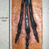
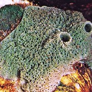
Freshwater sponges are widely distributed in well-oxygenated streams where they encrust on plant stems and old submerged pieces of wood. These sponges tend to be brownish or greenish and may resemble a bit of wrinkled scum pitted with pores. Two common North American freshwater genera are Spongilla and Myenia. Freshwater sponges are most commonly found in midsummer, although others may be more easily found in the fall. While freshwater sponges reproduce sexually, they may reappear commonly from gemmules. Freshwater sponges die in later autumn when the body disintegrates, leaving the asexually formed gemmules to overwinter and start the next year’s population.
In Arkansas, only seven species (all Demospongiae) of freshwater sponges have been documented: Anheteromeyenia argyrosperma (Potts); Dosilia radiospiculata (Mills); the Oneida sponge, Ephydatia mulleri (Lieberkühn); Lesser tendril sponge, Heteromeyenia latitenta (Potts); Trochospongilla horrida (Weltner); Trochospongilla leidyi (Bowerbank); and Spongilla fragilis (Leidy). It was not until 1936 that the first report of a freshwater sponge (T. horrida) was documented in Arkansas. Later, in 1951, two additional species were reported from the state: E. mulleri on a floating log from Savoy (Washington County) and T. leidyi from rocks at Eden’s Bluff (Washington County) and the Petit Jean River in Scott County. At the same time, T. horrida was found on rocks at the White River at Elkins (Washington County). In 1977, five species of freshwater sponges were reported from Beaver Lake in northwestern Arkansas, and in 2005, three species of sponges were reported in an abstract from DeGray Lake, including A. argyrosperma, H. latitenta, and T. horrida. Unknown species have been reported from Lake Ouachita. No further work has been done on the sponges of Arkansas due to a combination of a lack of trained workers, a changing taxonomy, and difficulty of finding these small invertebrates.
For additional information:
Annandale, N. “Fresh-water Sponges in the Collection of the United States National Museum. Part II. Specimens from North and South America.” Proceedings of the United States National Museum 37 (1909): 401–406.
———. “Fresh-water Sponges in the Collection of the United States National Museum. Part V. A New Genus Proposed, with Heteromeyenia radiospiculata Mills as Type.” Proceedings of the United States National Museum 40 (1911): 593–594.
Borchiellini, C., M. Manuel, E. Alivan, N. Boury-Esnault, J. Vacelet, and Y. LeParco. “Sponge Paraphyly and the Origin of Metazoa.” Journal of Evolutionary Biology 14 (2001): 171‒179.
Carter, H. J. “History and Classification of the Known Species of Spongilla.” Annals & Magazine of Natural History (Fifth Series) 7 (1881): 77–107.
Causey, David. “Freshwater Sponges in Arkansas.” Proceedings of the Arkansas Academy of Science 4 (1951): 89‒90. Online at: http://scholarworks.uark.edu/cgi/viewcontent.cgi?article=1117&context=jaas (accessed January 17, 2019).
Causey, David, and H. Eidson. “Trochospongilla horridus in Arkansas.” Science 84 (1936): 291.
Duncan, T. O. “Freshwater Sponges in Beaver Reservoir, Northwest Arkansas.” Southwestern Naturalist 22 (1977): 140.
Frost, T. M., H. M. Reiswig, and A. Ricciardi. “Porifera.” In Ecology and Classification of North American Freshwater Invertebrates, 2nd ed., edited by J. H. Thorp and A. P. Covich. New York: Academic Press, 2001.
Hooper, J., and R. W. M. van Soest, eds. A Guide to the Classification of Sponges. New York: Kluwer Academic/Plenum Publishers, 2002.
Mills, H. “A New Freshwater Sponge, Heteromeyenia radiospiculata n. sp.” Annals & Magazine of Natural History, Including Zoology, Botany, and Geology, Sixth Series 1 (1888): 313–314.
Pennak, Robert W. Fresh-Water Invertebrates of the United States: Protozoa to Mollusca, 3rd ed. New York: John Wiley & Sons, 1989.
Potts, E. “Contributions towards a Synopsis of the American Forms of Fresh Water Sponges with Descriptions of Those Named by Other Authors and From All Parts of the World.” Proceedings of the Academy of Natural Sciences of Philadelphia 39 (1887): 158–279.
———. “A New Form of Fresh-Water Sponge.” Proceedings of the Academy of Natural Sciences of Philadelphia 1881 (1881): 176.
Saul, T. P., M. R. Dare, and James A. Engman. “Freshwater Sponge Community Composition and Characteristics of Occurrence on Egeria densa in DeGray Lake, Arkansas, and a Report of Sponge Occurrence in Lake Ouachita, Arkansas [abstract].” Proceedings of the 89th annual meeting of the Arkansas Academy of Science, Hendrix College, Conway, Arkansas, 2005.
Smith, D. G. Pennak’s Freshwater Invertebrates of the United States: Porifera to Crustacea. New York: John Wiley and Sons, 2001.
Smith, F. “Distribution of Fresh Water Sponges in North America.” Bulletin of the Illinois State Natural History Survey 14 (1921): 9‒22.
World Porifera Database. http://www.marinespecies.org/porifera/porifera.php (accessed January 17, 2019).
Henry W. Robison
Sherwood, Arkansas
Chris T. McAllister
Eastern Oklahoma State University


Comments
No comments on this entry yet.